49 KiB
What is neuroscience?
Neuroscience is a field of scientific study that seeks to understand how the nervous system carries out its functions and what goes wrong when it doesn’t.

https://canvas.ucsc.edu/courses/46898
Note:
video of microetching 'self-reflection' project and info on how it was made by G. Dunn and B. Edwards
Welcome. This class will be an Introduction to Neuroscience–
Neuroscience is a field that by necessity integrates information and techniques from many other scientific disciplines— not just biological sciences like genetics, molecular biology, biochemistry, immunology, physiology. But also physics, engineering, computer science, psychology. And these days neuroscience is touching upon fields as varied as sociology, criminology, marketing, ethics, and the law. So what is Neuroscience? Neuroscience is fundamentally a field that...
And ultimately it is a field of science that seeks to understand how this lump of biological tissue siting inside our heads has evolved the capability of asking questions about its own nature and existence.
"carries out its functions"
--
Syllabus and text book


--
Site keyboard bindings
- Navigate: arrow keys and
spacebar - Overview toggle:
ooresc - Fullscreen:
f - Show notes:
s - Search toggle:
ctrl-shift-f - Menu toggle:
m - List keyboard shortcuts:
?
What are the nervous system’s functions?
- The nervous system organizes an individual’s interactions with the environment
- Its functions are dynamic and wide-ranging– extending to include all actions, thoughts, perceptions of the self.
Navigation.
Note:
What does the nervous system do? It organizes and controls an individuals interactions with the environment. It does this by processing current or past experiential information and making and executing behavioral decisions.
Therefore the brain’s functions are dynamic and vast, extending to include all thoughts, perceptions, and actions and the core of what it means for each of one us to be us–– consciousness and the mind. It is this complex lump of biological tissue, this emergent computational system that allows us humans to not only imagine the future, but to create it as well.
--
Neuroscience and the future of humankind?


Note:
Ever since the dawn of the industrial age in the mid 19th century and Jules Verne's 1865 novel 'From the Earth to the Moon' humans have been dreaming of the future, not just here but among the stars. And those futures can become reality like when the Apollo astronauts landed on the moon and acknowledged the inspiration that Verne's orig sci-fi novel had on many.
-
Neuroscience and its role for proper physiological function is going to play a role in many advances in health and technology for humankind now and far into the future--
-
To reach the stars we will need:
- robots, artificial intelligence, I. Asimov Philip K. Dick's 1968 novel 'Do Androids Dream of Electric Sheep'
- virtual reality, brain machine interfaces, James Cameron's Avatar
- medical tricorders, 1960s series Star Trek
- physiological stasis, cryopreservation, waking up the brain space after travel like Joe Haldeman's 1974 novel 'The Forever War' or the Ridley Scott's movie Aliens
The human brain and its limitless creativity has packed a bunch of computational power into this little device in our pocket. And yet this device is really just made up of lots of simple little semiconductive elements. So what is the atomic unit of our brains function and how is it structured to achieve our cognitive abilities and our consciousness? We will find the answers to some of these question in this course, but will also discover as is usually the case when looking into nature's secrets that we humbly know so little.
Or perhaps a discomforting future where maybe consciousness will be woven into some sort of singular virtual world like in the matrix or the cylons from Battlestar Galactica in ten or ten thousand years? Oh shoot wait, the metaverse is already here in 2021.
Think of virtual reality, can we solve the mismatches between sensory information and body positioning to get rid of the nausea associated with VR technology? Think of artifical intelligence and robotics
Traveling through space (well technically we are already traveling through space together on spaceship earth;) we will need to keep our bodies in working order to get wherever we are going-- will we know enough about brain function and neurolgical disease to fix things on the fly with a medical tricorder device like in Star Trek?
Can we read or even predict the minds of a suspect in a courtroom with a brain imaging device? Should we even want to do that?
We've dreamed up fantastical futures in shows like Star Trek and the Jetsons and dystopian ones in Blade Runner and the Terminator or even ones from the past (e.g "A long time ago in a galaxy far far away...")
Some of the things dreamed of are or will soon be present. Flying aeroplanes, personal landspeeders, rocket ships to distant planets, autonomous-automobiles.
- Edgar Rice Burroughs A Priestess of Mars: John Carter hurling thought waves.
- Do Androids Dream of Electric Sheep: Penfield mood organ (W. Penfield was an prominent canadian neuroscientist)
- So is the nervous system then a device for
- detecting physiological change?
- seeking pleasurable rewards? a dopamine machine?
- processing emotional secretions?
- navigating space-time?
Questions to keep in mind as we study neuroscience now and beyond
- What signals are produced by a nervous system? How?
- How are inputs from the external or internal environment transduced by the nervous system?
- How do sensor inputs combine with a self's existing world model to allow decisions and an output to be actuated?
- Structure? Function?
Note:
- Nervous
- relating to or affecting the nerves
- nervosus
- latin
- sinewy, vigorous
- nervus
- latin
- sinew
- sinew
- fibrous tissue linking bone or muscle to bone
- the parts of a structure, system, or thing that give it strength or bind it together
What are brains made of?
A glob of squishy jello?


Interacting sets of protoplasmic colloidal containers. Cells.
Note:
So what are brains made of? Anybody? Jello? What is this 1.5 kg or 3 lb human brain made of?
Yes it is soft and squishy but it is not just a gelanitous mass like jello. Thought jello is lots of collagen, and we are lots of collagen, including whaterver a brain is. Shown here is a section through a human brain. It is about 20 cm long and if we were to zoom in on a tiny part of it and use a special dye and microscope what we see is that the brain is made of cells. So this is a pyramidal neuron in from the cerebral cortex and its cell body is about 30-40µm in diameter.
(though jello is made of collagen...)
- colloid (wn, noun)
- (a mixture with properties between those of a solution and fine suspension)
- protoplasm (wn, noun)
- (the substance of a living cell (including cytoplasm and nucleus))
--
Elemental entities

O. Benfey's 1964 table, commons.wikimedia.org CC BY-SA 3.0
Note:
Based on Otto Theodor Benfey's spiral periodic table from 1964: https://en.wikipedia.org/wiki/Periodic_table#/media/File:Elementspiral_(polyatomic).svg
--
Sizes of some different entities
| object | diameter (millimeters) |
|---|---|
| dot, sand grain, paramecium | 1.0 |
| animal cell, muscle, bone, brain cells | 0.01 |
| bacteria, mitochondria, chloroplast | 0.001 |
| virus (influenza) | 0.0001 |
| protein | 0.000001 |
| water molecule | 0.000000275 |
Note:
Radius for sphere model protein:
- Rmin 1 <--> 5 nm
- Size 5 <--> 500kDa
- 1 angstrom = 0.1 nm
- 2.75 angstroms = 0.275 nm == H2O diameter
Brains consist of interconnected cells
- Camillo Golgi (Italy)– believed that cells in the brain were directly connected forming a continuous network (reticular theory).
- Santiago Ramon y Cajal (Spain)– Brains made up of single cells and communicate at specialized areas called synapses.
- Shared Nobel prize in 1906
Note:
Seems fairly obvious now. But wasn't in the 19th c. Cells widely accepted everywhere else in the 1830’s. But neuroscientists were the last to accept this right up until the turn of the 20th c.
Only after fundamental and rigorous work by these two scientists, C. Golgi and S. Ramon y Cajal in the late 19th c. did we come to appreciate comprised of individual cellular elements rather than a continous network or syncytium.
Golgi staining
Golgi staining: potassium chromate and silver nitrate (1873)


Note:
Golgi's drawing of hippocampus after performing his black potassum chromate and silver nitrate stain. Bottom is a zoomed in drawing of neurons and their connections in the hippocampal dentate gyrus.
The nervous system is not a syncytium
- syncytium: a mass of cytoplasm with many nuclei but no internal cell boundries
- reticulum: a fine network or netlike structure
- Camillo Golgi, Nobel Lecture December 11, 1906, The Neuron Doctrine- theory and facts:
"...Far from being able to accept the idea of the individuality and independence of each nerve element, I have never had reason, up to now, to give up the concept which I have always stressed, that nerve cells, instead of working individually, act together, so that we must think that several groups of elements exercise a cumulative effect on the peripheral organs through whole bundles of fibers."
Note:
Golgi drew the structure of the hippocampus as being all fused together into a reticulum, no free axon endings
Syncytiums are important in living organisms. From the placenta at the beginning of your existence to your multinucleated myocytes and osteocytes that make up your muscle and bones as you chase the Pacific Sun, syncytiums always play an important role.
The Neuron Doctrine
- Santiago Ramon y Cajal
- Neurons are cells. Each is an individual entity from an anatomic and embryologic view
- Function, however, involves the connection across spacetime between multiple entities
- Neurons have a polarity

Note:
Neurons in culture have specific endings. EM methods, dye filling experiments.
Heinrich Wilhelm Gottfried von Waldeyer-Hartz (6 October 1836 – 23 January 1921) was a German anatomist and conceived the word 'neuron'.
Golgi in his nobel lecture:
(3) The neuron is a physiological unit. This fundamental idea which Waldeyer expressed with perfect precision has been enlarged upon both from anatomical and functional sides with additional propositions, for example : The communication between neurons is only established by casual contact. There is scarcely any nervous tissue apart from the neurons; the neurons are also trophic units.
individual entitites. boxes within boxes. containers.
The Nobel Prize in Physiology or Medicine 1906
"in recognition of their work on the structure of the nervous system"

Camillo Golgi Pavia University Pavia, Italy

Santiago Ramón y Cajal Madrid University Madrid, Spain
Note:
How many neurons in a human brain?
- 100 thousand
- 10 million
- 100 million
- 1 billion
- 10 billion
- 100 billion
- 1 trillion
Note:
- in cerebral cortex humans generally have most neurons, where we have about 20 billion. Even compared to an elephant that has 3 times the number of overall neurons. Though some species of cetaceans (whales and dolphins) approach the number of our cortical neurons and recent research has shown that the long-finned pilot whale likely has more neurons in its cerebral cortex than we do.
100 billion sands grains could fit in a dump truck (~ 5 meters long).
d=1 #unit diam of sand grain as perfect sphere
#sphere fits in perfect cube with side length equal to d. Let d=1mm, then:
100e9 ** (1/3)

#so 4641 mm side cube or 4.6 m cube filled with uniformm 1 mm particles
4641**3
A billion hours ago our ancestors were living in the stone age A billion days ago no one walked on earth
Nervous system function is the product of genes and environment(s)
- Many genes are expressed in the brain, either during development or in the adult. It is the spatial and temporal regulation of these genes together with an organism's interaction with the environment that builds a nervous system.
- "nature
ORAND nurture"
Note:
- Neuroscience encompasses many fields: genetics, molecular and cell biology, developmental biology, physiology, physics, electrical engineering, computer science.
- not nature or nurture, nature and nurture
- language, learning to ride a bike
- clones, identical twins
Model the world in terms of entangled energy, entangled variances of polarized spatiotemporal change.
Genome size does not predict nervous system complexity
| organism | # of genes | # of base pairs | # of neurons | development time (young adult) |
|---|---|---|---|---|
| Caenorhabditis elegans (nematode) | ~19,000 | ~97 million | 302 | 8 hrs |
| Drosophila melanogaster (fruit fly) | ~15,000 | ~120 million | ~250,000 | 7–11 days |
| Danio rerio (zebrafish) | ~24,000 | ~1.5 billion | ~10,000,000 | 30 days |
| Xenopus tropicalis (frog) | ~21,000 | ~1.7 billion | ~16,000,000 | 4 months |
| Mouse | ~25,000 | ~3.5 billion | ~71,000,000 | 2-3 months |
| Human | ~20,000 | ~3.5 billion | ~100,000,000,000 | 18 years |
| African elephant | ~20,000 | ~3.1 billion | ~267,000,000,000 | 18 years |
Note:
Number of genes is not related to nervous system complexity or size. The nematode c. elegans has just 302 neurons, and yet its genome contains virtually as many genes as a humans. An african elephant brain weighs 3 times more than a human brain and has 3 times the number of neurons.
Even number of base pairs: Paris japonica (white, star like flower) has 150 billion base pairs of DNA (50x larger than that of a human haploid genome)
The largest brains are those of sperm whales, weighing about 8 kg (18 lb). An elephant's brain weighs just over 5 kg (11 lb), a bottlenose dolphin's 1.5 to 1.7 kg (3.3 to 3.7 lb), whereas a human brain is around 1.3 to 1.5 kg (2.9 to 3.3 lb). Brain size tends to vary according to body size.
| animal | n neurons | n synapse |
|---|---|---|
| roundworm | 302 | 7500 |
| jellyfish | 5600 | |
| sea slug | 18,000 | |
| amphioxus | 20,000 | |
| larval zebrafish | 100000 | |
| fruitfly | 250,000 | <10,000,000 |
| ant | 250,000 | |
| honey bee | 960,000 | |
| cockroach | 1,000,000 | |
| guppy | 4,300,000 | |
| frog | 16,000,000 | |
| zebra finch | 131,000,000 | |
| brown rat | 200,000,000 | 4.48x10^11 |
| red junglefowl | 221,00,000 | |
| ferret | 404,000,000 | |
| gray squirrel | 453,660,000 | |
| octopus | 500,000,000 |
sources for n neurons
- http://faculty.washington.edu/chudler/facts.html
- https://en.wikipedia.org/wiki/List_of_animals_by_number_of_neurons
E. Coli K-12: 4,639,221 bp, 4377 genes
Naegleria gruberi (unicellular free living eukaryote organism) 41 x 10^6 bp 15,727 genes
xenopus laevis | 3.1 billion bp | 16,948; 23,676 genes | ? neurons | 1 year | 1.3 mm egg
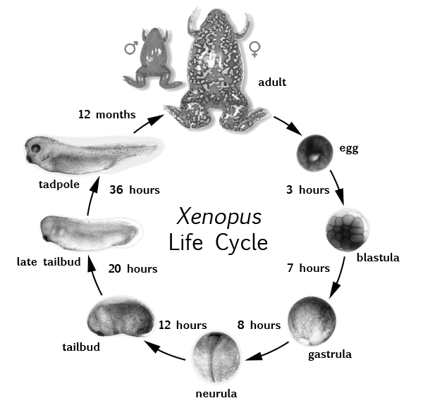
xenopus tropicalis | 1.7 billion bp | 21,634 genes | 16,000,000 neurons | 4 months | 0.8 mm egg
https://www.xenbase.org/anatomy/intro.do xenopus genome project http://viewer.shigen.info/xenopus/
- X. tropicalis develops faster with a higher optimal temperature (25-30°C vs 16-22°C), and has a diploid, smaller genome (instead of tetraploid) compared with X. laevis. Still has big eggs (0.8mm vs 1.2mm), large brood size (500-2000+) and large (but smaller) adults (4-5cm vs 10cm) as X. laevis
- gastrulation at 7-8hrs, tailbud at 20hrs and tadpole at 36hrs. Reproductive adults at 12mo.
- https://www.xenbase.org/anatomy/static/intro/xenopus_life_cycle_small.png
- xenopus hatchlings
- can detect light intensity (pineal gland, then with eyes a few days later) Foster and Roberts 1982)
- discriminate touch (skin is excitable with cardiac-like all or none APs for a primitive noxious stimuli response like Cnidarians (Mackie 1970). Body surface innervated by touch neurons to trigeminal gangli and spinal cord (Roberts 1980)
- lateral line neuromasts caudal to eyes for water current responses by swimming into them (Roberts 2009)
- have few thousand functioning central neurons (populations of 20-150) on each side [Roberts:2010]
[Roberts:2010]: Roberts A, Li WC, Soffe SR. How neurons generate behavior in a hatchling amphibian tadpole: an outline. Front Behav Neurosci. 2010 Jun 24;4:16. doi:10.3389/fnbeh.2010.00016. PMID: 20631854; PMCID: PMC2903309.
- Dictyostelium discoideum
- 34 Mb haploid genome, six chromosomes, 12500 genes
- soil dwelling amoeba, slime mold
- eukaryote transitioning between unicelular to multicellular slugs to fruiting body
- found in soil, moist leaf litter. diet is bacteria like E. Coli
- bacteria secretion of folic acid attracts the myxamoebae, which divide by mitosis during the vegetative stage why consuming bacteria
- mostly asexual lifecycle-- vegetative, aggregation, migration, culmination
- starvation during aggregation causes the myxamoebae to make glycoproteins that help cell-cell adhehsion and adenylyl cyclase which makes cAMP. cAMP works as a chemotactic signal, attracting neighboring amoebae then form a motile pseudoplasmodium, a slug up to 2-4mm long and 100000 cells
- https://upload.wikimedia.org/wikipedia/commons/thumb/3/3e/Cell-Shape-Dynamics-From-Waves-to-Migration-pcbi.1002392.s007.ogv/180px--Cell-Shape-Dynamics-From-Waves-to-Migration-pcbi.1002392.s007.ogv.jpg
- 5μm spores and amoebas to aggregated multicellular slugs of 1mm length
- sexual reproduction also possible. three mating types with three strains having different gene combinations that specify the three different sexes, can only mate with the two different sexes https://doi.org/10.1126%2Fscience.1197423 uhttps://pubmed.ncbi.nlm.nih.gov/21148389
- https://en.wikipedia.org/wiki/Dictyostelium_discoideum
- during chemotaxis, cAMP and amoeba movements occur every six minutes with amoebae moving toward concentration gradient for 60s before stopping. Oscillations in groups of cells results with propogating spiral waves of varying cAMP concentrations http://www.whydomath.org/Reading_Room_Material/ian_stewart/2000_11.html
- may exhibit food husbandry or 'farming' behavior! https://doi.org/10.1038%2Fnature09668
- Drosophila 7-11 days (28-34degs C)
- zebrafish 3-4 days juvenile swimming and visual behavior. young adult at 3 mo. full adult at 6 mo.
- genome sizes at http://www.biology-pages.info/G/GenomeSizes.html
What about cerebral cortical neuron number?
- generally humans thought to have highest neocortical neuron number (and therefore greatest amount of cortical links, inter-connects)
- but cetaceans approach and even rival humans in these measures... dolphins, killer whales, blue whale
- indeed a study in the past decade revealed the long-finned pilot whale as having more neocortical neurons than human.
- Globicephala melas; latin "globe" and greek kephale "head"
- up to 6.7 m long, 2300kg. second larges delphinid next to the Orcinus orca
- very social, small long term social units of 8-12 individual, aggregates of 100s to 1000s of individuals observed. usually small familial pods.
- gestation 12 - 16 months, calives are up to 2.0 m at birth and 75kg
- sex maturity for females and males at eight and 12 years respectively
- https://en.wikipedia.org/wiki/Long-finned_pilot_whale
https://en.wikipedia.org/wiki/List_of_animals_by_number_of_neurons#cite_note-80
African elephant 5,600,000,000 Pallium (cortex) Loxodonta africana1
Chimpanzee 7,400,000,000 Pallium (cortex) Pan troglodytes2
Bottlenose dolphin 12,700,000,000 Pallium (cortex) Tursiops truncatus3
Human 21,000,000,000 Pallium (cortex) Homo sapiens4
Blue whale 15,000,000,000 Pallium (cortex) Balaenoptera musculus3
Long-finned pilot whale 37,200,000,000 Pallium (cortex) Globicephala melas5
Killer whale 43,100,000,000 Pallium (cortex) Orcinus orca3
There are many brain-specific and non-brain specific genes expressed in the nervous system
| tissue | Number of expressed genes |
|---|---|
| brain only | ~6000 |
| brain & all other tissues | ~8000 |
| other tissues only | ~6000 |
| total: 20000 |
Note:
Out of those 20000 genes, there are many expressed genes that are common between the nervous system and other tissues, however there is also a substantial fraction that are expressed specifically in the nervous system
A single mutation can lead to dramatic brain size defects
Mutation in a spindle pole gene call ASPM1 (altered mitosis during brain development)

Bond:2002, see also Neuroscience 5e Fig. 1.1
Note:
Now mutations in single genes in the right place in our genome can cause drastic effects on the formation of our brain’s wiring.
For example, shown here is a person with a mutation in ASPM1 a protein used to make spindle poles for mitotic stem cells during embryonic development.
But most single gene mutations do not cause such drastic effects, with a more subtle and complex set of genetic and environmental risk factors causing neurological disease, similar to and probably exceeding the complex etiology of cancer.
2cm scale bar. left 13yr old female patient. right 11 yr old control.
Glial cells
- Glia
- greek for 'glue'
- outnumber neurons 10-50 fold (higher mammals)
- structural support for neurons
- remove debris and maintain a functional nervous system environment
Note:
Now there are two basic cell types in the nervous system, neurons and glia. We will revisit neurons more in a few minutes and will be talking all about their function over the ensuing lectures but first lets touch briefly on some of the types of glial cells and their known functions.
Up to 90% of brain cells in mammals.
During evolution the glia/neuron ratio basically follows a power relation ship 6 y(x) = kx^n where on a log-log plot k is the intercept and n is the slope. Some of this original comparative estimates of glia/neuron ratios among animals was performed by Friede (1954)
Perhaps only 10% of cells in invertebrates like drosophila.
Other model organisms also have nervous system support cells like glia-- C. elegans has just 56 total 'glial cells'. They fall into three major populations (24 sheath cells, 26 socket cells, and 6 GLR). The 6 GLR cells are mesodermally derived.

- afrotheria
- african species
- shrews, west indian manatees, elephants, moles
Types of glia
- Astrocytes– Support cells of the CNS, most numerous type of glia
- Microglia- CNS macrophages. Act as phagocytes, mobilized after infection, injury, or disease
- Oligodendrocytes– Myelin producing cells of the CNS
- Schwann cells– Myelin producing cells of the PNS
- Satellite cells– Support cells of the PNS


Note:
Satellite glial cells are glial cells that cover the surface of nerve cell bodies in sensory, sympathetic and parasympathetic ganglia.
--
Astrocytes
- Restricted to CNS
- Maintain a proper chemical environment
- Deliver metabolic support to neurons from blood vessels
- Help maintain the blood-brain barrier
- Neurochemical recycling at synapses


Note:
Astrocytes are star shaped, hence their name.
Astrocytes are your pizza delivery persons for neurons. They are also like your mom, constantly upkeeping your room or synapses as is the case for neurons.
They are the direct decendents of the mother stem cells that give rise to the neurons and glia of the nervous system.
Devasting diseases of astrocyte function include brain cancer with gliomas like glioblastomas typicaly being comprised of astrocytes gone wild. It is also thought that some childhoold epilepsies may originate from altered astrocyte function.
blood brain barrier-- control entry of neurotransmitters and hormones into the brain
areas of the brain without a blood-brain barrier (from Table 32-2 Basic Neurochemistry 6e):
Pituitary gland Median eminence Area postrema Preoptic recess Paraphysis Pineal gland Endothelium of the choroid plexus
There is a positive relationship between lipid solubility and brain uptake of chemical compounds:
- permeability of lipid soluble compounds is rapid (ethanol, nicotine, diazepam, THC)
- polar molecules (e.g. glycine and catecholamines) enter slowly across BBB
- gases and volatile anesthetics diffuse rapidly into the brain
blood—brain barrier permeability of CO2 greatly exceeds that of H+ thus pH of brain interstitial fluid reflect pCO2 rather than blood pH. Therefore a patient with metabolic acidosis may be brain alkalotic at the same time.
glucose is primary energy substrate of the brain. Nearly all oxygen consumption for the brain. GLUT-1 glucose transporters highly enriched in brain capillary endothelial cells. Since glucose is a polar substrate, this transporter facilitates its transport across the BBB.
Neutral L-amino acids enter the brain as rapidly as glucose (Phenylalanine, leucine, tyrosine, isoleucine, valine, tryptophan, methionine, histidine and l-dihydroxy- phenylalanine (l-DOPA))
water enters rapidly through diffusion.

See also review by 7 for info on energy dynamics between astrocytes-neurons.
--
Oligodendrocytes
- Insulate axons in CNS by wrapping in myelin sheaths. Myelination is essential for electrical signal propagation
- Each cell can myelinate multiple axons


Note:
Multiple sclerosis or MS is an example of a devastating CNS disease characterized by degeneration of the myelin sheaths.
--
Schwann cells
- Myelinate axons in peripheral nervous system (PNS)
- One axon per cell

Note:
Discovered by German scientist Theodore Schwann. In 1839 he actually stated that all animal tissues are made of cells.
A number of other demylinating diseases other than MS that involve schwann cell dysfunction. Charcot–Marie–Tooth disease (CMT), Guillain–Barré syndrome.
Neurons
- Main signaling unit of the nervous system
- Polarized– have dendrites and axons and a direction for information flow
- Communicate by electricity– usually using action potentials.
- Tremendous range of different cell types– categorized by morphology, molecular identity and physiological activity.
Note:
Now let’s think about the cell type most responsible for the brain’s business of biological computation— the neuron.
It is the...
Which of the following cell structures are found in neurons?
- DNA
- RNA
- Nucleus
- ER
- Mitochondria
- Microtubules
- Golgi
- Cell division machinery
Note:
--
Cell body (soma) of a neuron

Note:
- soma is another word for cell body
- the processes extending away from the cell body, the dendrites and axons are filled with cytoskeletal support like microtubles and actin filaments. Provide shape and structure to the neuron and are important during development of processes. Neurodegenerative diseases like alzheimers often affect components of the cytoskeleton (microtubles or actin filaments)
Neurons have a functional polarity
- Incoming information arrives and is integrated among the dendrites and cell body
- The integrated information is then relayed along the axon to the next neuron via synapses
Note:
Polarity is everywhere and is everything, in physics... and biology!
- electric dipole moments of molecules
- earth's magnetic poles
- electromagnetic waves
- DNA (5'-->3')
- mitotic cells
- apical-basal orientation of cells within tissues
- animal embryos and neural tube
from oxford dict,
- polar
- directly opposite in character or tendency
- polarity
- the relative orientation of poles; the direction of a magnetic or electric field
- the tendency of organisms or parts to develop with distinct anterior or posterior ends, or to grow or orient in a particular direction
Structures of a neuron
- Cell body (soma)– metabolic center of the cell, contains the nucleus.
- Dendrites– receive incoming signals from other nerve cells
- Axon– carries signals to other neurons
- Axon hillock– initiates action potentials
- Synapse– site at which two neurons communicate
- Synaptic cleft– area between pre and post-synaptic cell
Note:
Neuron processes: dendrites
- Dendrites
- Extensively branching from the cell body
- Transmit electrical signals (graded potentials) toward the cell body
- Function as receptive sites for other neurons
Note:
--
Dendritic spines


Note:
- 2 billion transistors in an iphone6.
- 100 billion neurons, each receiving up to 10000 synaptic connections
- quadrillion synapses, 10^15 in our nervous system
False color of the dendrite of one neuron near an axon from another neuron from an EM image
- semiconductors 22nm to 14nm (half distance between nodes on the array)
- synaptic vesicles, avg diameter of 40nm 8
- diameter of neurofilament 10nm 8
- thickness of neuronal membrane 5 nm 8
- synaptic cleft distance 20-40nm 8
- internodal length 150-1500µm 8
- dendritic spine membrane area in rat striatum-- 0.5µm^2 == 0.4µm radius == 0.8µm diameter 9
- neck diameter 0.15µm 9
- spine density 40 spines/10nm 9
Neuron processes: axons
- Axons (nerve fibers)
- Each neuron has only one, but it can branch
- Neurofilaments, actin microfilaments, and microtubules
- Provide structural strength along length of axon
- Axonal transport of biochemical substances
- Carry neuronal electrical signals (action potentials) away from the cell body
Note:
?chalkboard
- Branches along length are infrequent. End is called terminal bouton or axonal arbors
- Aid in the transport of substances to and from the cell body
- Impulse generator and conductor
- Axon collaterals
- Multiple branches at end of axon
- Terminal branches
- End in knobs called axon terminals (also called terminal boutons)
Neurons are classified in different ways
- Morphology: unipolar, bipolar, and multipolar
- Function: sensory neurons, motor neurons, and interneurons
- Neurotransmitter expression: excitatory, inhibitory, dopaminergic, etc.
Note:
- similar classes of cells and morphologies and neuronal shapes found in the human nervous system as in other animals. People have looked hard but there doesn't appear to be any class of cell that is unique to humans or higher mammals-- i.e. no unique neuron subtype that makes us human. We'll talk alot about the neurochemical differences that underly different types of neurons later in the course and their different functional properties.
--
Example morphologies– cerebellar neurons

Note:
--
Example morphologies– cortical neurons
- Pyramidal neurons– multipolar neurons that contain both apical and basal dendrites. Also contain one axon eminating from cell body
- Most common excitatory neuron in the cerebral cortex

.svg)


Note:
--
Example morphologies– retinal neurons


Note:
todo: need Coombs et al., 2006 figures...
Structure of a sensory neuron (afferent)
Function of an afferent neuron is to carry information from the sensory periphery towards the central nervous system.

Note:
Afferent- term meaning to send information from periphery to the CNS or to brain
Structure of a motor neuron (efferent)
Function of an efferent neuron is to carry information towards the muscles for bringing about behavior.

Note:
Efferent sends info to muscles
Affect vs effect
Neurons communicate with electrical pulses
- Axons project great distances
- Use action potentials to transmit information
- Neuronal interactions ('functional connections') occur at synapses
- separated by small amounts of space– the synaptic cleft (~40 nm)
- Action potential causes release of neurotransmitter that is received by post-synaptic cells
Note:
What is electricity? It's energy. It's variance. From the flow of electric charge across a conducting medium.
--
Inter-neuronal signaling occurs at synapses

Note:
We will be going into synapse structure and function in much detail later in the class, but just to complete our introduction to basic anatomical details of neurons this figure illustrates...
Neuron signals: action potentials
- Nerve impulse (action potential or 'spike')
- Neuron receives and sends signals
- Generated at the initial segment of the axon
- Conducted along the axon
- Releases neurotransmitters at axon terminals
- Neurotransmitters – excite or inhibit neurons
Note:
We will be discussing the nature of basic unit of nervous conduction, the action potential or impulse in great detail in ensuing lectures.
Properties of the action potential
- rapid
- transient
- all or none
- self-regenerating
- can go long distances. 5 m in a giraffe
- highly stereotyped
- discrimination is based on patterns of firing
Note:
-
15 m if you're a branchiosaurus
-
rate coding
-
phase coding
Neural Circuits
- Neurons don’t function in isolation, they are organized into circuits that process specific kinds of information
- Direction of information flow is important for understanding the function of a circuit
- Afferent neurons– carry information toward the brain
- Efferent– carry info from the brain
Note:
Example of a simple circuit: stretch (myotatic) reflex
The "knee-jerk response" is a simple reflex circuit.

Note:
Muscle lengthens, stretching muscle spindle (sensory ending), leading to incr alpha motor neuron activity and causing same muscle group to contract. Works to maintain muscle length.
- stretch tendon and sensory recpetors in leg extensor muscle
- sensory neuron synapse with and excites motor neuron in spinal cord
- sensory neuron also excites spinal interneuron
- interneuron synapse inhibits motor neuron to flexor muscles
- motor neuron cducts APs to synapse on extensor muscle fibers causing contraction
- flexor muscle relaxes because its motor neurons activity has been reduced
- leg extends
Ways to measure neural activity
- Extracellular recording– an electrode is placed near a neuron. Measures action potentials. Useful for detecting patterns of activity.
- Intracellular recording– an electrode is placed inside a neuron-can measure smaller graded potential changes. Useful for isolating responses to single inputs.
Note:
You might have the anatomy skills of Cajal or Golgi and you know there is this reflex you're studying and you've seen the morphologies of hundreds of cells along this pathway, but what is the cells function during this behavior, how do you monitor that?
Extracellularly recorded responses underlying the stretch reflex

Note:
These ticks are spikes or action potentials recorded extracelluarly. Since the electrode tip is placed close to the neurons cell membrane, the electrode can pick up signals as they pass by. A little bit like someone wiretapping your phone line.
We will come back to this reflex circuit in greater detail time and again as we go through this course.
And really, the basic logic of this circuit and variants of it is replicated all over the brain and teasing apart all the types of cells, their response properties, and their functional interactions or connections with one another for all types of different sensory and motor behavior is the grand challenge, beauty, and fun of modern and future neuroscience.
-
Herculano-Houzel S, Avelino-de-Souza K, Neves K, Porfírio J, Messeder D, Mattos Feijó L, et al. The elephant brain in numbers. Front Neuroanat. (2014). 8:46. doi:10.3389/fnana.2014.00046 ↩︎
-
Collins CE, Turner EC, Sawyer EK, Reed JL, Young NA, Flaherty DK, et al. Cortical cell and neuron density estimates in one chimpanzee hemisphere. Proc Natl Acad Sci U S A. (2016). 113:740–5. doi:10.1073/pnas.1524208113 ↩︎
-
Herculano-Houzel S. Longevity and sexual maturity vary across species with number of cortical neurons, and humans are no exception. J Comp Neurol. (2019). 527:1689–1705. doi:10.1002/cne.24564 ↩︎
-
Herculano-Houzel S, Catania K, Manger PR, Kaas JH. Mammalian brains are made of these: A dataset of the numbers and densities of neuronal and nonneuronal cells in the brain of glires, primates, scandentia, eulipotyphlans, afrotherians and artiodactyls, and their relationship with body mass. Brain Behav Evol. (2015). 86:145–63. doi:10.1159/000437413 ↩︎
-
Mortensen HS, Pakkenberg B, Dam M, Dietz R, Sonne C, Mikkelsen B, et al. Quantitative relationships in delphinid neocortex. Front Neuroanat. (2014). 8:132. doi:10.3389/fnana.2014.00132 ↩︎
-
Bélanger, M., Allaman, I., and Magistretti, P. J. (2011). Brain energy metabolism: focus on astrocyte-neuron metabolic cooperation, Cell Metab, 14(6), 724-38 ↩︎